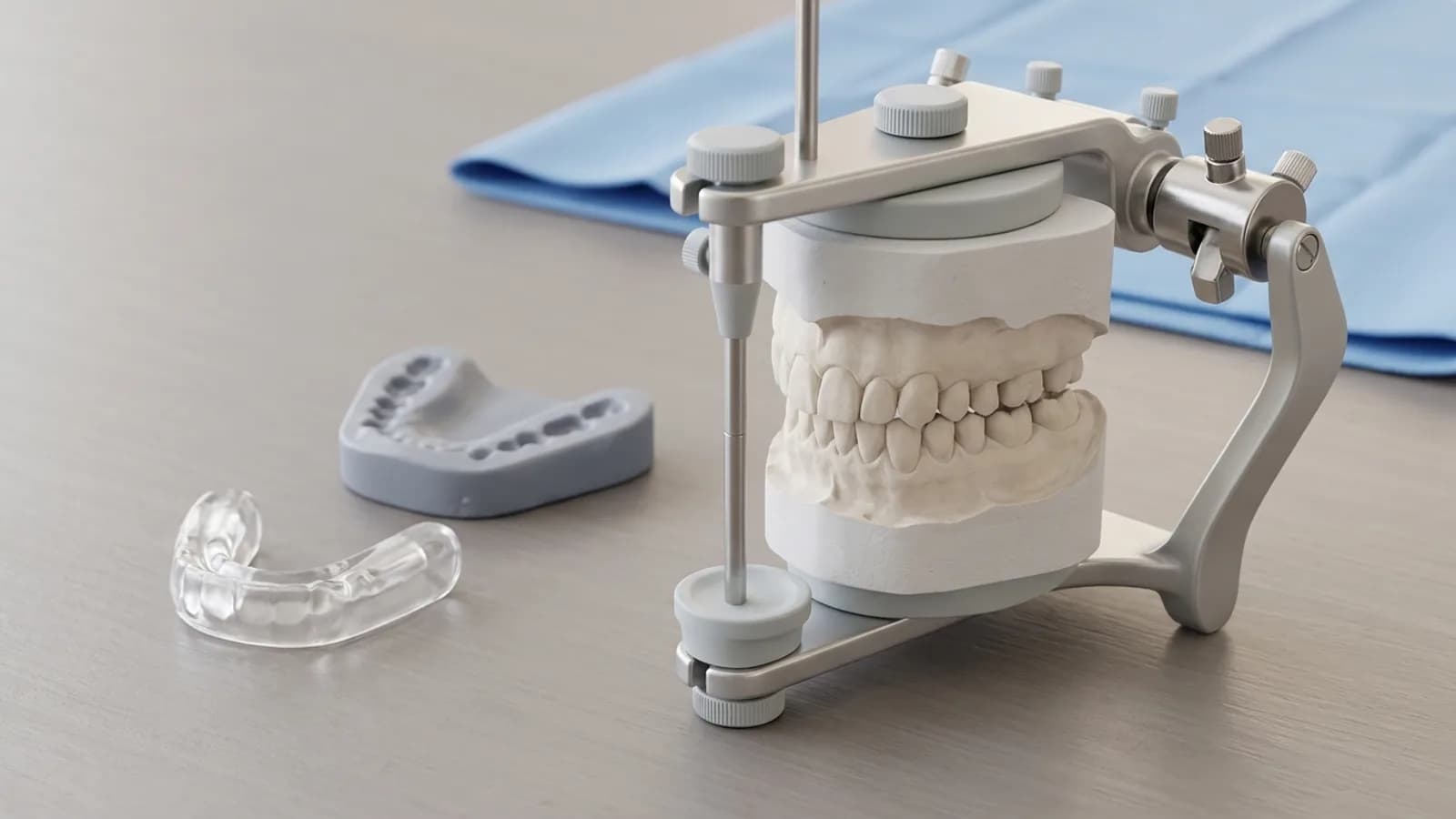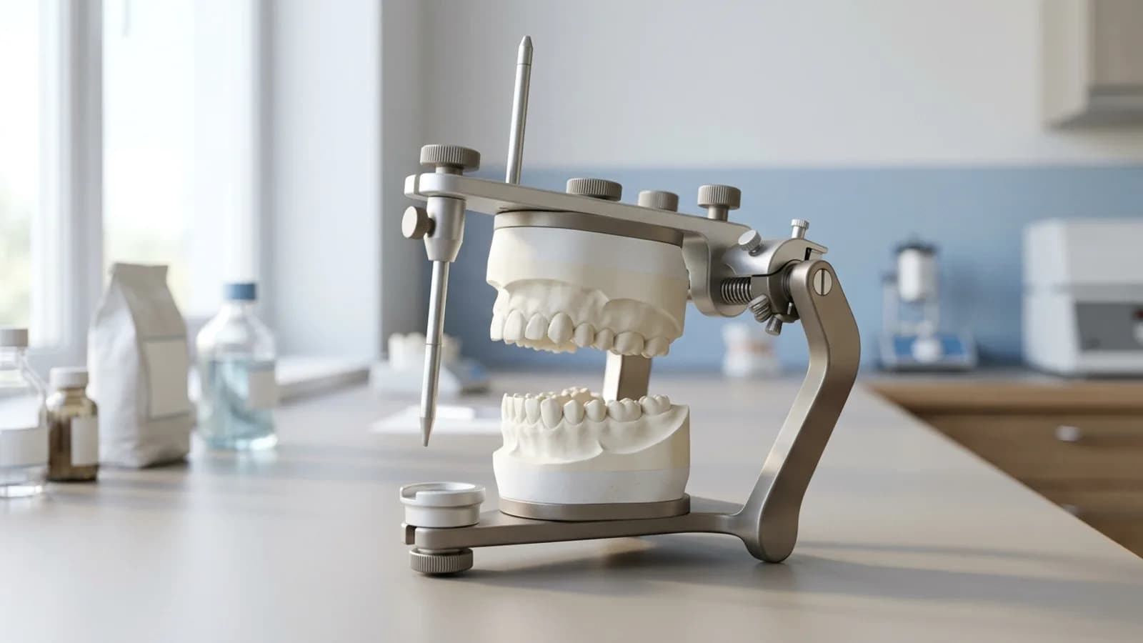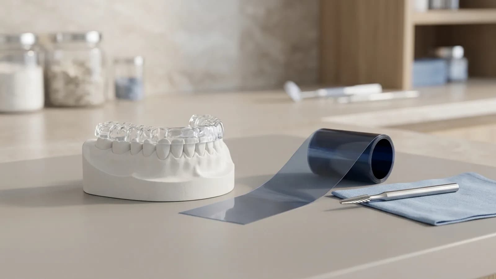
An occlusal splint is the most widely used reversible treatment for temporomandibular disorders (TMD) and a standard tool before complex restorative work. Whether it helps depends less on the brand of appliance than on the diagnosis, the jaw registration and how carefully the splint is adjusted. This article sets out when splint therapy is indicated, what the trials actually show, and a chairside protocol for delivering and adjusting a hard stabilisation splint of the Michigan type.
When is an occlusal splint indicated?
Start with a diagnosis, not with the appliance. The Diagnostic Criteria for Temporomandibular Disorders (DC/TMD) give a validated framework for separating pain-related diagnoses such as myalgia and arthralgia from intra-articular disorders such as disc displacement (Schiffman et al. 2014). A short screening at the check-up identifies which patients need a full functional examination at all.
Indications fall into two groups.
Functional indications
- myalgia of the masticatory muscles
- arthralgia and painful joint clicking
- bruxism, mainly to protect teeth and restorations
- traumatic occlusion, for example a high new restoration
- differential diagnosis: if symptoms settle under an occlusally neutral splint, the masticatory system is probably involved
Restorative indications
- before a systematic occlusal adjustment
- severe tooth wear before rehabilitation
- before extensive fixed restorations when the mandibular position is to be changed
- as a trial of an increased vertical dimension in collapsed or worn dentitions; see vertical dimension of occlusion
For painful TMD the treatment goals are modest: less pain, adequate mouth opening, adequate chewing function and removal of contributing factors. The splint is non-invasive and fully reversible, which is its main advantage.
What the evidence says about occlusal splint therapy
The literature is more nuanced than many patient leaflets suggest.
Placebo-controlled trials support the stabilisation splint. Ekberg et al. randomised 60 patients with mainly myogenous TMD to a stabilisation splint or a non-occluding control splint. After 10 weeks the splint group reported overall improvement significantly more often, with greater reductions in daily pain and in the number of tender muscles (Ekberg et al. 2003). The benefit persisted at 6 and 12 months (Ekberg and Nilner 2004). In mainly arthrogenous TMD the same group found an advantage for the stabilisation splint, although the control splint also had an effect (Ekberg et al. 1998).
A meta-analysis of 33 randomised trials found short-term benefits for pain, muscle tenderness and mouth opening, particularly in muscular TMD; in the long term the effect was similar to other treatments. Continuous wear during the day was associated with better results (Kuzmanovic Pficer et al. 2017).
The systematic review by Riley et al. (2020, 37 trials) is more sceptical. When all TMD subtypes were pooled, no reduction in pain could be shown, and the certainty of evidence was very low. No trial measured whether splints reduce tooth wear in bruxism.
A network meta-analysis of 48 trials ranked a hard stabilisation splint combined with counselling among the most effective options for both myogenous and arthrogenous TMD, with moderate to very low certainty of evidence (Al-Moraissi et al. 2020).
The practical conclusion: use the splint as one part of a multimodal plan, not as the whole treatment. As early as 1998 Dao and Lavigne asked whether splints are "crutches" and recommended them as an adjunct to pain management rather than a definitive treatment. In bruxism the realistic goal is protection of teeth and restorations.
Splint types: what you need to know
| Splint type | Principle | Typical use |
|---|---|---|
| Hard stabilisation splint (Michigan type) | full coverage, flat occlusal surface, canine guidance | standard for myogenous and arthrogenous TMD, bruxism, pre-restorative phase |
| Mandibular stabilisation splint | same principle, guidance built on the splint | alternative to the maxillary splint |
| Soft splint | resilient material, cannot be adjusted precisely | short-term at most |
| Anterior repositioning splint | holds the mandible forward | selected disc displacement cases, time-limited |
| Anterior bite stop (jig) | anterior contact only | deprogramming, registration, short-term |
The Michigan splint described by Ramfjord and Ash is the best documented design: hard acrylic, full coverage of the maxillary arch, a flat occlusal surface contacted by the opposing cusp tips, canine guidance in excursions and minimal opening of the vertical dimension (Ramfjord and Ash 1994).
A note on repositioning: there is no evidence to support restoring a dentition extensively in a protruded mandibular position. Prosthodontic treatment is not an initial TMD therapy and should only follow, on restorative grounds, once reversible treatment has relieved pain and dysfunction (De Boever et al. 2000). In an anterior position the vertical dimension also changes with the jaw position, which makes a stable centric stop hard to achieve.
Maxillary or mandibular splint?
Both work if the contacts and guidance are right. Comparative trials showing a clear advantage for either arch are scarce, so the criteria below are practical considerations rather than study findings.
| Favours a mandibular splint | Favours a maxillary splint |
|---|---|
| a long, flat guidance is easy to build | the anterior guidance should become steeper |
| usually less effect on speech and daytime wear | little room in the lower arch, e.g. a large tongue |
| contacts easier to adjust chairside | bonded retainer in the lower arch |
| cheek biting (lateral shields possible) | |
| marked overjet |
Also ask: has the patient worn a splint before, and did they tolerate it? Is restorative, orthodontic (aligner) or provisional work planned in one arch during the splint phase? Is splinting of mobile teeth desirable? Switching arches occasionally improves compliance. German-speaking readers will find a patient-oriented discussion of upper versus lower splints in our article Aufbissschiene, aber richtig (in German).
Hands-on dental courses
Courses for dentists in Munich, several of them taught in English.
See all coursesRegistration and amount of opening
A splint is only as good as the jaw relation it is made in. An anterior jig or leaf gauge separates the posterior teeth and helps to deprogramme the muscles before the record is taken. The step-by-step technique is covered in centric relation registration with an anterior jig.
How much should you open?
- With a substantial loss of vertical dimension: to the estimated future restorative height, so the splint doubles as a trial of the new vertical dimension.
- In all other cases: as little as possible, but enough for the splint to stay flat over the last molar with adequate material thickness there. The minimum thickness of the splint material is given by its manufacturer.
The proportion of rotation versus translation on opening and the posterior cusp inclination both influence how much anterior opening is needed.
Delivery and adjustment step by step
Get the fit right before touching the occlusion: the splint should seat stably and passively, without pressure points or excessive play. If it does not, reline it first.
- Check the fit: seat the splint and look for rocking, flexing and pressure points. If needed, sandblast the fitting surface, apply a bonding agent and reline with light-cured resin after isolating the teeth.
- Static occlusion: with the patient relaxed, let them close without force, guiding the mandible at most with a light touch at the chin and never pushing it. The target is one contact for each supporting cusp of the opposing arch on a flat surface, with no indentations and no occlusal anatomy.
- Use fine foil: a 12 µm occlusion foil marks precisely and resists tearing (manufacturer information, Coltène HANEL OccluFoil+). Thicker articulating papers tend to mark broader areas, which makes individual contacts harder to judge.
- Dynamic occlusion: canine guidance should be smooth, long enough and, if in doubt, slightly flatter rather than steeper. No posterior interferences in laterotrusion or protrusion. The principles behind this are discussed in canine guidance and the occlusion concept.
- Check the opposing canine: a heavily worn canine tip may need a small composite addition before the splint can provide clear guidance.
- Adjust in very small steps, re-marking each time.
- Smooth and polish.
- Record every adjustment and addition in the notes.
If too much has been removed, or a contact is missing after the jaw position has changed, add material: sandblast the area, apply bonding agent, isolate the opposing teeth with petroleum jelly, place a clear filled composite, ask the patient to close gently, tack-cure, complete curing outside the mouth, then adjust as above.
Reviews until the contacts are stable
The splint is worn at least at night and, where possible, during the day, consistent with the association between longer wear and better outcomes (Kuzmanovic Pficer et al. 2017).
There are no evidence-based review intervals. Reviews are usually close together at first and spaced out as the contacts stabilise, with a shorter interval again after major corrections. Ask patients to have worn the splint before each review and to bring it to every dental appointment.
The adjustment phase is complete when the splint contacts no longer change from one review to the next. For muscular pain, counselling on parafunction and self-observation belongs in the plan, since splint plus counselling ranked among the most effective options in the network meta-analysis (Al-Moraissi et al. 2020). Many clinicians also involve a physiotherapist.
Digital splints
Splints can be scanned in the opened position, designed digitally and milled or printed. The clinical protocol stays the same: a CAD/CAM splint must also fit without tension and be adjusted and reviewed by the same criteria. For printed splints, follow the resin manufacturer's instructions on post-curing and cleaning.
Summary
Successful occlusal splint therapy starts with a diagnosis, uses a hard, full-coverage stabilisation splint with flat contacts and canine guidance, and depends on meticulous adjustment at short intervals. The evidence supports the splint as part of a multimodal approach, not as a cure-all. The full sequence from screening and splint to the restorative phase is taught hands-on in our VDO and occlusion training course for dentists.
Frequently asked questions
How long does occlusal splint therapy take?
There is no fixed duration. The adjustment phase ends when the contacts no longer change between reviews, which usually takes several weeks. Meta-analytic data show the clearest benefit in the short term. After that, the course of symptoms decides whether the splint stays as a protective night appliance or a restorative phase follows.
Should the splint be made for the upper or the lower arch?
Both work if the splint provides flat posterior contacts and smooth canine guidance, and trials have not shown a clear advantage for either arch. In practice, a mandibular splint makes a flat guidance easy to build and usually affects speech less. A maxillary splint suits a marked overjet, a bonded lower retainer or little room in the lower arch.
Is a soft splint an alternative to a hard Michigan splint?
Not for therapy that relies on controlled occlusion. Soft splints cannot be adjusted precisely, so flat contacts and defined canine guidance are hard to achieve. In the network meta-analysis by Al-Moraissi et al. the evidence for soft splints was of very low quality. The hard, full-coverage stabilisation splint remains the standard.
Does an occlusal splint stop bruxism?
No. A splint protects teeth and restorations from bruxism forces but does not stop the activity itself. Whether it measurably reduces tooth wear has not been studied adequately; Riley et al. (2020) found no trials that measured it. In bruxism with muscle pain, a stabilisation splint is still a reasonable part of management.
How thick should a stabilisation splint be?
As thin as possible and as thick as necessary. Open the registration just enough for the splint to stay flat over the last molar while keeping the minimum material thickness specified by the splint material's manufacturer. With substantial loss of vertical dimension, register at the planned restorative height instead, so the splint tests the new vertical dimension before any teeth are restored.